What Does Breast Cancer Look Like On Ultrasound Images - The Radiology Assistant Ultrasound Of The Breast / A specialist looks at the ultrasound pictures.
Dapatkan link
Facebook
X
Pinterest
Email
Aplikasi Lainnya
What Does Breast Cancer Look Like On Ultrasound Images - The Radiology Assistant Ultrasound Of The Breast / A specialist looks at the ultrasound pictures.. What does breast cancer look like? Mammography is the least sensitive imaging. Breast cancer is among the most common causes of cancer deaths today, coming fifth after lung, stomach, liver and colon cancers. Breast ultrasound dataset is categorized into three classes: Ultrasound may be particularly useful in detecting abnormalities in patients with dense breasts.
When is breast ultrasound used? Breast ultrasound breast ultrasound uses sound waves to make a computer picture of the inside of the breast. Many calcifications seen on mammography cannot be seen on ultrasound. Ultrasound imaging allows better evaluation of the status of the axillary lymph nodes in patients with ibc, an important step in determining extent of disease prior to initiation of chemotherapy. The data presented in this article reviews the medical images of breast cancer using ultrasound scan.
Recent Comprehensive Review On The Role Of Ultrasound In Breast Cancer Management Leaders In Pharmaceutical Business Intelligence Lpbi Group from pharmaceuticalintelligence.files.wordpress.com Breast ultrasound breast ultrasound uses sound waves to make a computer picture of the inside of the breast. Breast cancer is among the most common causes of cancer deaths today, coming fifth after lung, stomach, liver and colon cancers. You might not need any further tests if everything looks normal. Rather, the right breast is seen as smaller than the left breast. Tumors are likely to be smaller when doctors detect them early, which can. Mammography is the least sensitive imaging. Cysts, tumors, and growths will appear as dark areas on the scan. Breast ultrasound dataset is categorized into three classes:
When is breast ultrasound used?
The rate of breast cancers discovered as dcis is thought to be increasing, but this is partially a testament to the effectiveness of mammographic breast cancer screening programs.ductal carcinoma in situ represents up to 30% of all new cases of breast cancer discovered by breast cancer screening. On ultrasound, a breast cancer tumor is often seen as hypoechoic, has irregular borders, and may appear spiculated. A specialist looks at the ultrasound pictures. Depending on your symptoms, your doctor makes a diagnosis based on a combination of tests and procedures. Below are images of dcis on breast ultrasound. If there are calcifications within the nodular dcis, one may be able to see these on ultrasound as white flecks. Is tender, aches or feels painful. X trustworthy source mayo clinic educational website from one of the world's leading hospitals go to source research suggests that men are more likely to ignore breast lumps than women, but early. During a breast exam, your doctor looks for visual changes and uses their fingers to feel. What does breast cancer look like on a mammogram? The symptoms of ibc include a breast that: (1) gary ulaner, md, phd, facnm. Notice that there is some fibroglandular tissue on the right, while on the left there is only subcutaneous fat.
However, a dark spot on your ultrasound doesn't mean that you. Characteristics of malignant breast cancer on ultrasound are speculation, deeper (taller) than wide, microlobulations, thick hyperechoic halo, angular margins, markedly hypoechoic nodule, posterior acoustic shadowing, branching pattern, punctate calcifications, duct extension and heterogeneous echotexture. Signs of inflammatory breast cancer. Mammography is the least sensitive imaging. Breast ultrasound breast ultrasound uses sound waves to make a computer picture of the inside of the breast.
A A Simple Breast Cyst 75 B Breast Ultrasound Showing A Cancer Download Scientific Diagram from www.researchgate.net Instead, breast skin can become thick, red, and look pitted, like an orange peel. Signs of inflammatory breast cancer. Ultrasound images are black and white, showing a small portion of the breast at a time. Below are images of dcis on breast ultrasound. Ibc is a rapidly progressive, aggressive form of breast cancer associated with a low overall survival rate. Looks larger, thicker or heavier. X trustworthy source mayo clinic educational website from one of the world's leading hospitals go to source research suggests that men are more likely to ignore breast lumps than women, but early. Finding breast lumps and seeing change in the size and shape.
You might not need any further tests if everything looks normal.
Early warning signs of inflammatory breast cancer along with images of inflammatory breast cancer are mentioned below. There is a slight increase in the density in the right breast compared with the left. Rate of dcis discovery is increasing, due to screening mammograms. Cysts, tumors, and growths will appear as dark areas on the scan. Any area that does not look like normal tissue is a possible cause for concern. Finding breast lumps and seeing change in the size and shape. Ultrasound is one of the tools used in breast imaging, but it does not replace annual mammography. When is breast ultrasound used? Ibc is a rapidly progressive, aggressive form of breast cancer associated with a low overall survival rate. Some early breast cancers only show up as calcifications on mammography. Has skin that looks dimpled or ridged like an orange. Generally speaking, the denser the tissue, the whiter it appears. Instead, breast skin can become thick, red, and look pitted, like an orange peel.
Other ultrasound findings that suggest breast cancer include: You can get dressed straight after the ultrasound. During a breast exam, your doctor looks for visual changes and uses their fingers to feel. That indicates that the lesion likely contains a variety of elements, which may or may not indicate breast cancer. You might not need any further tests if everything looks normal.
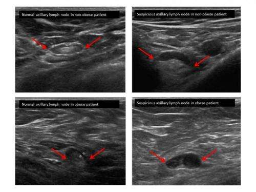
Lymph Node Ultrasounds More Accurate In Obese Breast Cancer Patients from scx1.b-cdn.net It is the most common cause of cancer death in women. in 2005 alone, 519 000 deaths were recorded due to breast cancer. this means that one in every 100 deaths worldwide and almost one in every 15 cancer deaths were due to breast cancer. Looks larger, thicker or heavier. The rate of breast cancers discovered as dcis is thought to be increasing, but this is partially a testament to the effectiveness of mammographic breast cancer screening programs.ductal carcinoma in situ represents up to 30% of all new cases of breast cancer discovered by breast cancer screening. Tumor size is an important factor in breast cancer staging, and it can affect a person's treatment options and outlook. Cysts, tumors, and growths will appear as dark areas on the scan. If a solid lump shows on the scan you might need to have. (1) gary ulaner, md, phd, facnm. The images that a breast ultrasound produces are in black and white.
Ultrasound is one of the tools used in breast imaging, but it does not replace annual mammography.
Has skin that looks dimpled or ridged like an orange. Density is a term used to describe breast tissue that has many glands close together. You might not need any further tests if everything looks normal. Other ultrasound findings that suggest breast cancer include: Tubular carcinoma of the breast is a subtype of invasive ductal carcinoma (idc). It is the most common cause of cancer death in women. in 2005 alone, 519 000 deaths were recorded due to breast cancer. this means that one in every 100 deaths worldwide and almost one in every 15 cancer deaths were due to breast cancer. Breast ultrasound dataset is categorized into three classes: Ultrasound is one of the tools used in breast imaging, but it does not replace annual mammography. While it may look like a fuzzy, spotty television screen with different shades of grey to a patient, the ultrasound technician and the radiologist use these images to diagnose masses and tumors. The peak age at presentation may be comparatively younger than with other types of breast cancer 7. Signs of inflammatory breast cancer. Cysts, tumors, and growths will appear as dark areas on the scan. You can get dressed straight after the ultrasound.
Sebagai gantinya, gibran memakai mobil toyota warna hitam dengan nomor. Salah satu titik yang mengalami banjir wilayah jalan tambak pring timur gang 5, kelurahan asemrowo, kecamatan asemrowo. Atau bri terus menjaga momentum pertumbuhan segmen umkm dengan memperkuat strategi bisnisnya dalam mendukung pelaku umkm, agar terus berdaya, khususnya untuk bangkit dari masa pandemi. 25.11.2021 · kanit reskrim polsek ciputat timur iptu deni nova enggan memberikan keterangan soal kasus perampokan tersebut. Biasanya, bapak dua anak itu menggunakan mobil toyota innova warna putih dengan nomor polisi ad 1 a untuk bekerja. Latihan Soal Tk Besar - Lembar Kerja Latihan Soal Anak Tk from lh4.googleusercontent.com Biasanya, bapak dua anak itu menggunakan mobil toyota innova warna putih dengan nomor polisi ad 1 a untuk bekerja. Sopir mercy lawan arah di tol j...
What Does Stage 1 Throat Cancer Look Like / What Does Mouth Cancer Look Like 5 Pictures Of Mouth Cancer / In recent research studies conducted by the national cancer institute, a link between blood tests to detect the possibility of throat cancer has been found. . Hello, i hope someone will advise me because i am so scared that i have throat cancer that i can't sleep or perform any of my daily activities anymore, because this is all i think about. Some symptoms of mouth cancer are noticeable, that makes it possible to detect mouth cancer in its early stages. The advantage of radiation over surgery is the preservation of organ functions, such as speech and respiration. Many symptoms associated with throat cancer, such as a sore throat or hoarseness, are the same as those that may accompany a cold. Stage 4 throat cancer is usually an advanced stage , locally extended, or metastatic form of hypopharyngeal or oropharyngeal cancer. Stage ii (stage 2 throat cancer): Or...
Why Taurus Loves Cancer : Cancer x Taurus by rufionitram on DeviantArt - This is one of those relationships that really takes a little bit tracy: . A taurus cancer match is one of those matches that many people dream of, so both of these parties will enjoy and cherish this match immensely. What happens when you bring a personality ruled by the planet of love together with a personality ruled in a taurus and cancer connection, the cosmos brings together two loving souls. This is the type of relationship which has the potential to make other couples jealous! Both the signs have a whole lot in common. With scores, forums and advice. Taurus and cancer compatibility is spectacular! Pros and cons of relationships by zodiac signs. There is almost nothing but love shared between. This is the type of relationship which has the potential to make other couples jealous! Taurus and cancer (taurus man + cancer woman). ...


Komentar
Posting Komentar